
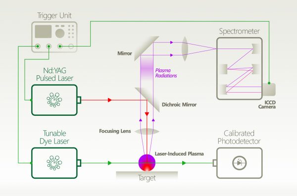
 Principle of Laser-Induced Plasma Spectroscopy Principle of Laser-Induced Plasma SpectroscopyLaser-induced plasma has been used for different diagnostic and technological applications as detection, thin film deposition, and elemental identification. Laser-Induced Plasma Spectroscopy ( LIPS), or sometimes called Laser-Induced Breakdown (LIBS) or Laser Spark Spectroscopy (LSS) is a powerful tool for rapid in-situ analyses of solid, liquid or gaseous samples. Some examples include on-line monitoring in the steel industry, control of materials and leakages in power plants, environmental analyses (soils, waters), analyses of objects related to Cultural Heritage, explosive identification in the protection against terrorism and planetary exploration (surface characterization).
Pulsed laser deposition is a thin film deposition (specifically a PVD) technique, where a high power pulsed laser beam is focused inside a vacuum chamber to strike a target of the deposit material. Conceptually and experimentally, pulsed laser ablation is an extremely simple technique; probably the simplest among all thin-film growth techniques.
With progress in short-pulse laser techniques, newly generated plasma applications appear. Extreme ultraviolet (EUV) sources based on laser-produced plasmas (LPP) emitting at a wavelength of tens of nm are currently being developed with tremendous effort for the next generation of semiconductor microlithography. Besides high-power sources for high volume manufacturing, compact EUV sources of lower average power are also needed for metrology purposes, for example, for mask inspection, actinic material testing, or optics and sensor characterization. Moreover, tabletop LPP sources are also employed to generate soft x-ray (SXR) radiation for microscopy or absorption spectroscopy in the water window spectral range (2.2–4.4 nm).
The availability of synchrotron x-ray and laser-plasma x-ray sources have revolutionized the capabilities of time-resolved x-ray diffraction investigations, the development of streak camera x-ray detectors, static and streak mode charge-coupled device (CCD) x-ray area detector techniques, and specialized scintillation detection schemes.
Effects of pressure and substrate temperature on the growth of Al-doped ZnO films by pulsed laser deposition
Related products: NL300 series
Optical coherence tomography (OCT) with 2 nm axial resolution using a compact laser plasma soft X-ray source
Related products: NL300 series
XUV spectra of 2nd transition row elements: identification of 3d–4p and 3d–4f transition arrays
Related products:
Initiation of vacuum insulator surface high-voltage flashover with electrons produced by laser illumination
Related products: NL300 series
Peculiarity of convergence of shock wave generated by underwater electrical explosion of ring-shaped wire
Related products: NL300 series
Emission properties of ns and ps laser-induced soft x-ray sources using pulsed gas jets
Related products: SL230 series APL2100 series
 Principle of Laser Flash Photolysis Principle of Laser Flash PhotolysisPhotodissociation, photolysis, or photodecomposition is a chemical reaction in which a chemical compound is broken down by photons. It is defined as the interaction of one or more photons with one target molecule. Photolysis plays an important role in photosynthesis, during which it produces energy by splitting water molecules into gaseous oxygen and hydrogen ions. This part of photosynthesis occurs in the granum of a chloroplast where light is absorbed by chlorophyll. This reacts with water and splits the oxygen and hydrogen molecules apart.
Laser Flash Photolysis is a technique for studying transient chemical and biological species generated by the short intense light pulse from a nanosecond/picosecond/femtosecond pulsed laser source (pump pulse). This intense light pulse creates short lived photo-excited intermediates such as excited states, radicals and ions. All these intermediates are generated in concentrations large enough for chemical and physical interaction to occur and for direct observation of the associated temporally changing absorption characteristics. Typically the absorption of light by the sample is recorded within short time intervals (by a so-called test or probe pulses) to monitor relaxation or reaction processes initiated by the pump pulse. Usage of Optical Parametric Oscillators (OPO) opens new possibilities in spectroscopic experiments.
Electronic spectroscopy and nanocalorimetry of hydrated magnesium ions [Mg(H2O)n]+, n = 20–70: spontaneous formation of a hydrated electron?
Related products: NT230 series NT242 series NT342 series
Infrared spectroscopy of O˙− and OH− in water clusters: evidence for fast interconversion between O˙− and OH˙OH−
Related products: NT270 series NT370 series
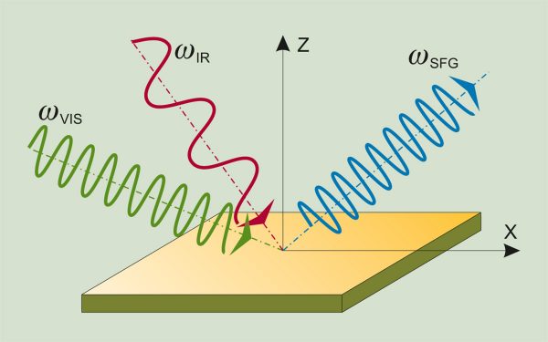 Principle of Sum Frequency Generation Vibrational Spectroscopy Principle of Sum Frequency Generation Vibrational SpectroscopySum frequency generation vibrational spectroscopy (SFG-VS) is used to characterize vibrational bonds of molecules at surfaces or interfaces. SFG spectroscopy is particularly attractive because of molecular specificity and intrinsic interfacial sensitivity. Surface sensitivity of the technique arises from the fact that within the electric dipole approximation the nonlinear generation of the sum-frequency (SF) signal from the overlapped visible and infrared beams is forbidden in the media of randomly oriented molecules or in the centrosymmetric media but is allowed at the interface where inversion symmetry is broken. Molecular specificity emerges from the ability to record vibrational spectrum.
In SFG-VS measurements, carried out by employing SFG spectromer, a pulsed tunable infrared IR (ωIR) laser beam is mixed with a visible VIS (ωVIS) beam to produce an output at the sum frequency (ωSFG = ωIR + ωVIS). SFG signal is generated in visible spectral range, so it can be efficiently measured using sensitive detectors (PMT or CCD).
Hydrogen bonding interactions of H2O and SiOH on a boroaluminosilicate glass corroded in aqueous solution
Related products: SFG Spectrometer
Reconfiguration of interfacial energy band structure for high-performance inverted structure perovskite solar cells
Related products: SFG Spectrometer
Segregation of an amine component in a model epoxy resin at a copper interface
Related products: SFG Spectrometer
High-performance graphdiyne-based electrochemical actuators
Related products: SFG Spectrometer
Sum Frequency Generation Vibrational Spectroscopy for Characterization of Buried Polymer Interfaces
Related products: SFG Spectrometer
A structural and temporal study of the surfactants behenyltrimethylammonium methosulfate and behenyltrimethylammonium chloride adsorbed at air/water and air/glass interfaces using sum frequency generation spectroscopy
Related products: SFG Spectrometer PL2230 series PGx01 series
Discovery of Cellulose Surface Layer Conformation by Nonlinear Vibrational Spectroscopy
Related products: SFG Spectrometer
Sum frequency generation vibrational spectroscopy (SFG-VS) for complex molecular surfaces and interfaces: Spectral lineshape measurement and analysis plus some controversial issues
Related products: SFG Spectrometer
The complex nature of calcium cation interactions with phospholipid bilayers
Related products: SFG Spectrometer
Platelet-adhesion behavior synchronized with surface rearrangement in a film of poly(methyl methacrylate) terminated with elemental blocks
Related products: SFG Spectrometer
Alkanethiols as Inhibitors for the Atmospheric Corrosion of Copper Induced by Formic Acid: Effect of Chain Length
Related products: SFG Spectrometer
Simultaneous measurement of magnitude and phase in interferometric sum-frequency vibrational spectroscopy
Related products: SFG Spectrometer
Phase measurement in nondegenerate three-wave mixing spectroscopy
Related products: SFG Spectrometer
Probing the Orientation and Conformation of α-Helix and β-Strand Model Peptides on Self-Assembled Monolayers Using Sum Frequency Generation and NEXAFS Spectroscopy
Related products: SFG Spectrometer PL2230 series PGx01 series
Study of self-assembled triethoxysilane thin films made by casting neat reagents in ambient atmosphere
Related products: SFG Spectrometer PL2230 series
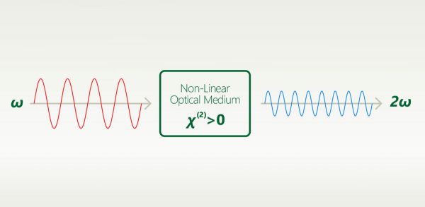 Principle of Second Harmonic Generation (SHG) Spectroscopy Principle of Second Harmonic Generation (SHG) SpectroscopySecond harmonic generation (SHG) is a second order nonlinear optical effect where two photons of frequency ω are converted to one photon of frequency 2ω. SHG is allowed only in media without inversion symmetry. This optical method it is non﹊nvasive, can be applied in situ, and can provide real time resolution.
SHG measurements provide information about: surface coverage, molecular orientation, adsorbtion-desorbtion processes, and reactions at interfaces. SHG has the ability to detect low concentrations of analytes, such as proteins, peptides, and small molecules, due to its high sensitivity, and the second harmonic response can be enhanced through the use of target molecules that are resonant with the incident (ω) and/or second harmonic (2ω) frequencies.
SHG microscopy allows for selective probing of a non-centrosymmetric area of sample. This type of nonlinear optical microscope was first used to observe ferroelectric domains and has been applied to various specimens including the biological samples to date. Imaging of the endogenous SHG of biological tissue can be utilized for the selective observation of filament systems in tissues such as collagen, myosin, and microtubules, which exhibit a polar structure. It has been reported that, by imaging exogenous SHG of the membrane, sensitive detection of membrane damage could be realized using the SHG microscope.
FemtoLux 3 laser for the rapid wide-field second harmonic generation microscopy
Related products: FemtoLux series
 Principle of Pump-Probe Spectroscopy Principle of Pump-Probe SpectroscopyUltrafast spectroscopy is based on the use of light pulses that have shorter temporal duration compared to the underlying dynamics of the system. Shorter pulse- better time resolution, longer pulse- better spectral resolution. Two pulses separated by time delay are used. First pulse excites (electronic and/or vibrational levels, ionises, make Coulombic explosions, creates plasma, etc.) sample, second delayed pulse probes- checks what happened at the certain time moment. Dynamics of investigated system is recorded by changing delay between pulses. Various experimental techniques are employed to record time-resolved signals: for example transient absorption, four-wave mixing, sum frequency generation. Pump and probe spectral range can vary from UV or even X-ray to far infrared.
The advantage of pump-probe spectroscopy is direct investigation of dynamics. For example: excitation relaxation, energy transfer, photochemical reactions dynamics and movement of particles, structural changes. Tunability of pump and probe pulses opens two dimensional pump-probe spectroscopy where is possible to obtain temporally resolved energy map of system which shows separated and coupled states.
Near infrared light induced plasmonic hot hole transfer at a nano-heterointerface
Related products: PL2210 series
Capturing an initial intermediate during the P450nor enzymatic reaction using time-resolved XFEL crystallography and caged-substrate
Related products: NT230 series
Luminescence upconversion in colloidal double quantum dots
Related products: NT342 series
Photogeneration and reactions of benzhydryl cations and radicals: A complex sequence of mechanisms from femtoseconds to microseconds
Related products: NT230 series NT242 series NT342 series
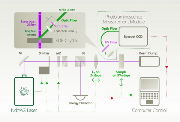 Principle of Solid-phase Photoluminescence Spectroscopy Principle of Solid-phase Photoluminescence SpectroscopyTime-resolved Photoluminescence Spectroscopy (TRPL) is a contactless method to characterize recombination and transport in solid materials. Measuring TRPL requires exciting luminescence from a sample with a pulsed light source and then measuring the subsequent decay in photoluminescence as a function of time. Most experiments excite the sample with a pulsed laser source and detect the PL with a photodiode, streak camera, or photomultiplier tube set up for upconversion or single-photon counting. The system response time, wavelength range and sensitivity vary widely for each configuration.
It is possible to apply the general methodology of time-resolved photoluminescence for lifetime imaging of the charge carrier dynamics. Nonradiative surface recombination at the boundaries of a semiconductor device can be a major factor limiting the efficiency in light-emitting and laser diodes (LEDs and LDs), photovoltaic cells, and photodetectors. Therefore, the effective lifetime is a crucial parameter to obtain solar cells with a high conversion ratio.
Photoluminescence microscopy also is a powerful optical method for the study of crystal defects in semiconductors and organometallic complexes, with important applications in the manufacturing process of nanostructures, optoelectronic devices and solar cell systems.
The detrimental effect of AlGaN barrier quality on carrier dynamics in AlGaN/GaN interface
Related products:
Superlinear Photoluminescence by Ultrafast Laser Pulses in Dielectric Matrices with Metal Nanoclusters
Related products:
Near infrared emission properties of Er doped cubic sesquioxides in the second/third biological windows
Related products: NT342 series
Ultra-sensitive mid-infrared emission spectrometer with sub-ns temporal resolution
Related products: PL2230 series PGx11 series
Probing the pathways of free charge generation in organic bulk heterojunction solar cells
Related products:
Quenching of the red Mn4+ luminescence in Mn4+-doped fluoride LED phosphors
Related products: NT342 series
Nanoscale insights into doping behavior, particle size and surface effects in trivalent metal doped SnO2
Related products: NT342 series
Non-Poissonian photon statistics from macroscopic photon cutting materials
Related products: NT342 series
Regio- and conformational isomerization critical to design of efficient thermally-activated delayed fluorescence emitters
Related products:
Revealing the spin–vibronic coupling mechanism of thermally activated delayed fluorescence
Related products:
Nile blue shows its true colors in gas-phase absorption and luminescence ion spectroscopy
Related products: NT342 series
Aerobic photoreactivity of synthetic eumelanins and pheomelanins: generation of singlet oxygen and superoxide anion
Related products: NT242 series
A cylindrical quadrupole ion trap in combination with an electrospray ion source for gas-phase luminescence and absorption spectroscopy
Related products: NT230 series NT242 series NT342 series
Roles of reactive oxygen species in UVA﹊nduced oxidation of 5,6ヾihydroxyindole�2ヽarboxylic acid﹎elanin as studied by differential spectrophotometric method
Related products: NT242 series
Room-temperature InP distributed feedback laser array directly grown on silicon
Related products:
Multi-photon quantum cutting in Gd2O2S:Tm3+ to enhance the photo-response of solar cells
Related products: NT342 series
Luminescence upconversion in colloidal double quantum dots
Related products: NT342 series
Study of GaN : Eu3+ Thin Films Deposited by Metallorganic Vapor-Phase Epitaxy
Related products: NT342 series
 Principle of Gas-phase Ion Luminescence Spectroscopy Principle of Gas-phase Ion Luminescence SpectroscopyGas-phase ion luminescence spectroscopy is used to determine intrinsic electronic transition energies in the absence of a disturbing environment. Large ions produced by electrospray ionization are stored and mass-selected in a cylindrical Paul trap. Here they are irradiated by light from a 20-Hz pulsed tunable wavelelength laser from EKSPLA followed by detection of the emitted photons. The 20-Hz repetition rate of the tunable laser allows for mass selection in between every irradiation event, which implies that there is no fluorescence contribution from ion impurities. The tunability of the laser makes it possible to photo-excite a variety of dyes that absorb at different wavelengths. The figure shows spectra of oxazine dye cations that display emissions in a range from 500 nm to 750 nm. The technique can also be used to study three-dimensional structures of peptides and nucleic acids in the gas phase based on Förster Resonance Energy Transfer (FRET). The biomolecules are labeled with donor-acceptor dye pairs such as rhodamines 575 and 640. Structure information is provided as the efficiency of energy transfer depends on the distance between the donor and acceptor to the inverse power of six.
Luminescence Spectroscopy of Rhodamine Homodimer Dications in Vacuo Reveals Strong Dye〥ye Interactions
Related products: NT342 series
Gas-phase Ion Spectroscopy of Flexible and Nonflexible Nitrophenolates: Effect of Locking the Two Phenyl Units in 4’-nitro-[1,1’-biphenyl]-4-olate by a Bridging Atom
Related products: NT342 series
Sibling rivalry: intrinsic luminescence from two xanthene dye monoanions, resorufin and fluorescein, provides evidence for excited-state proton transfer in the latter
Related products: NT342 series
Luminescence spectroscopy of chalcogen substituted rhodamine cations in vacuo
Related products: NT342 series
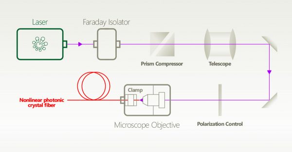 Principle of Supercontinuum Generation Principle of Supercontinuum GenerationSupercontinuum “White Light Lasers” have become a well-established turn-key fiber-laser technology addressing a wide range of applications from biomedical imaging to optical device characterization. They add value due to their unique combination of optical parameters, including an extremely wide spectral coverage from 400 nm to 2400 nm, several W of optical output power, and focus down to the diffraction limit of a perfect Gaussian beam.
The majority of commercial supercontinuum lasers are fully fiber-based systems consisting of a modelocked fiber oscillator as the master seed laser, providing ps pulses at ∼1064 nm and repetition rates in the tens of MHz regime. Injected into a fiber amplifier giving rise to high peak power, and finally a few meters of a specially designed index-guiding PCF with suitable dispersion landscape. The supercontinuum source, when combined with a tunable spectral filter, transforms into a widely tunable laser, making it a versatile laser tool for a wide range of applications.
Mid-infrared, super-flat, supercontinuum generation covering the 2–5 μm spectral band using a fluoroindate fibre pumped with picosecond pulses
Related products: PL2210 series PGx11 series
On-chip visible-to-infrared supercontinuum generation with more than 495 THz spectral bandwidth
Related products: LightWire FFS
 Principle of Time-Resolved Photoconductivity Principle of Time-Resolved PhotoconductivityTime-resolved photoconductivity (TRMC) are key techniques used to perform the contactless determination of carrier density, transport, trapping, and recombination parameters in charge transport materials such as organic semiconductors and dyes, inorganic semiconductors, and metal-insulator composites, which find use in conductive inks, thin-film transistors, light-emitting diodes, photocatalysts, and photovoltaics.
The behavior of photoconductivity with photon energy, light intensity and temperature, and its time evolution and frequency dependence, can reveal a great deal about carrier generation, transport and recombination processes. Many of these processes now have a sound theoretical basis and so it is possible, with due caution, to use photoconductivity as a diagnostic tool in the study of new electronic materials and devices.
The importance of relativistic effects on two-photon absorption spectra in metal halide perovskites
Related products: NT342 series
Charge carrier transport in polycrystalline CH3NH3PbI3 perovskite thin films in a lateral direction characterized by time-of-flight photoconductivity
Related products: NT342 series
Evidence of enhanced photocurrent response in corannulene films
Related products:
Investigation into the Advantages of Pure Perovskite Film without PbI2 for High Performance Solar Cell
Related products: NT342 series
Flexible non-volatile optical memory thin-film transistor device with over 256 distinct levels based on an organic bicomponent blend
Related products: NT342 series
Competition between recombination and extraction of free charges determines the fill factor of organic solar cells
Related products: NT242 series
Transparent Organic Photodetector using a Near-Infrared Absorbing Cyanine Dye
Related products: NT342 series
Efficient charge generation by relaxed charge-transfer states at organic interfaces
Related products: NT242 series
Time-of-flight mobility of charge carriers in position-dependent electric field between coplanar electrodes
Related products:
A novel method for direct nondestructive diagnosis of caries affected tooth surfaces by laser multiphoton ionization
Related products: NT342 series
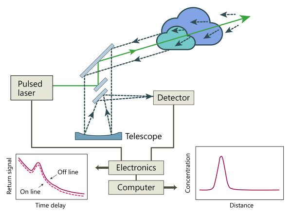 LIDAR operating principle LIDAR operating principleLIDAR is an acronym for “LIght Detection And Ranging”. LIDAR sends out short laser pulses into the atmosphere, where all along its path, the light is scattered by small particles, aerosols, and molecules of the air and is collected by telescope for analysis. Due to the constant velocity of light, time is related to the scatter’s distance, therefore, the spatial information is retrieved along the beam path. LIDAR uses ultraviolet, visible, or near infrared light to image objects. It can target a wide range of materials, including non-metallic objects, rocks, rain, chemical compounds, aerosols, pollutants, clouds, and even single molecules. LIDAR especially helps in those cases where access with conventional methods is troublesome.
 Z-scan operating principle Z-scan operating principleIn nonlinear optics, z-scan technique is used to measure the non-linear index n2 (Kerr nonlinearity) and the non-linear absorption coefficient Δα via the “closed” and “open” methods to measure both real and imaginary components of the nonlinear refractive index.
For measuring the real part of the nonlinear refractive index, the z-scan setup is used in its closed-aperture form. The sample is typically placed in the focal plane of the lens, and then moved along the z axis, defined by the Rayleigh length. In this form, since the nonlinear material reacts like a weak z-dependent lens, the far-field aperture makes it possible to detect small beam distortions in the original beam. Since the focusing power of this weak nonlinear lens depends on the nonlinear refractive index, it is possible to extract its value by analyzing the z-dependent data acquired by the detector and by interpreting them using an appropriate theory.
For measurements of the imaginary part of the nonlinear refractive index, or the nonlinear absorption coefficient, the z-scan setup is used in its open-aperture form. In open-aperture measurements, the far-field aperture is removed and the whole signal is measured by the detector. By measuring the whole signal, the beam small distortions become insignificant and the z-dependent signal variation is due to the nonlinear absorption entirely.
The main cause of non-linear absorption is two-photon absorption. Due to high pulse intensity and cost effectiveness,picosecond high energy lasers are the most appropriate choice for z-scan measurements.
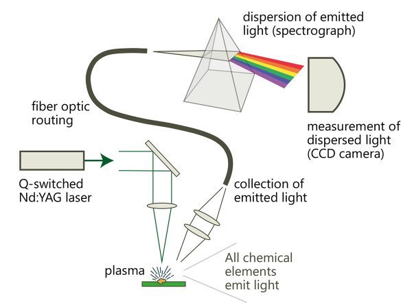 LIBS operating principle LIBS operating principleLaser-induced breakdown spectroscopy (LIBS) utilizes a high intensity short laser pulse to convert a very small amount of target material to plasma for optical analysis of the spectra. LIBS can be used on solid, liquid, or gas samples, and, depending on the spectrograph and detector, can detect all elements. LIBS is non-contact, so it can be used in a wide variety of environments, including remote analysis and micro-sampling.
When coupled with appropriate optics and stages, elemental maps of a surface can be created. Multiple LIBS scans can effectively resolve material composition throughout the volume, building a full three dimensional elemental map.
Adjusting the catalytic properties of cobalt ferrite nanoparticles by pulsed laser fragmentation in water with defined energy dose
Related products: Atlantic series
Overview of the application of laser-based techniques in plasma-wall interaction research program at IFPiLM
Related products:
Laser Induced Breakdown Spectroscopy and Applications Toward Thin Film Analysis
Related products: NL230 series NL300 series
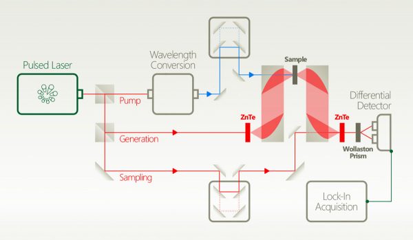 Principle of Terahertz Spectroscopy Principle of Terahertz SpectroscopyTerahertz time-domain spectroscopy (THz-TDS) probes inter-molecular interactions within solid materials. THz-TDS covers the spectral region of 0.1-10 THz which is a low energy and non-ionizing region of the electromagnetic spectrum. THz-TDS is a powerful technique for material characterization and process control and has several distinct advantages for use in spectroscopy. It can give the amplitude and phase information of the sample simultaneously. Many materials are transparent at terahertz wavelengths, and this radiation is safe for biological tissue being non-ionizing (as opposed to X-rays). It has been used for contact-free conductivity measurements of metals, semiconductors, 2D materials, and superconductors. Furthermore, THz-TDS has been used to identify chemical components such as amino acids, peptides, pharmaceuticals, and explosives, which makes it particularly valuable for fundamental science, security, and medical applications.
Terahertz Spectroscopy for Gastrointestinal Cancer Diagnosis
Related products: LightWire FFS
Green light stimulates terahertz emission from mesocrystal microspheres
Related products:
|


